
 back to blog
Multi-scale segmentation and surface fitting for measuring 3D macular holes
Selected photos from the paper below:
Click to go to full publication on IEEE Xplore.
Abstract
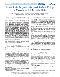
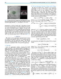
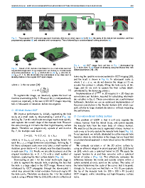
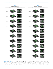
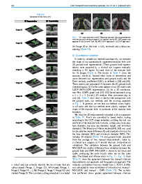
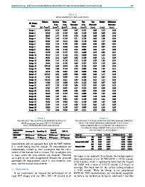
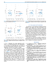
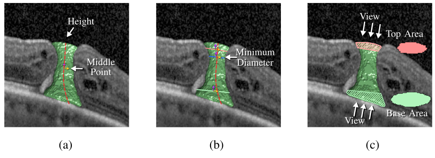
Macular holes are blinding conditions, where a hole develops in the central part of retina, resulting in reduced central vision. The prognosis and treatment options are related to a number of variables, including the macular hole size and shape. High-resolution spectral domain optical coherence tomography allows precise imaging of the macular hole geometry in three dimensions, but the measurement of these by human observers is time-consuming and prone to high inter- and intra-observer variability, being characteristically measured in 2-D rather than 3-D. We introduce several novel techniques to automatically retrieve accurate 3-D measurements of the macular hole, including: surface area, base area, base diameter, top area, top diameter, height, and minimum diameter. Specifically, we introduce a multi-scale 3-D level set segmentation approach based on a state-of-the-art level set method, and we introduce novel curvature-based cutting and 3-D measurement procedures. The algorithm is fully automatic, and we validate our extracted measurements both qualitatively and quantitatively, where our results show the method to be robust across a variety of scenarios. Our automated processes are considered a significant contribution for clinical applications.
Overview
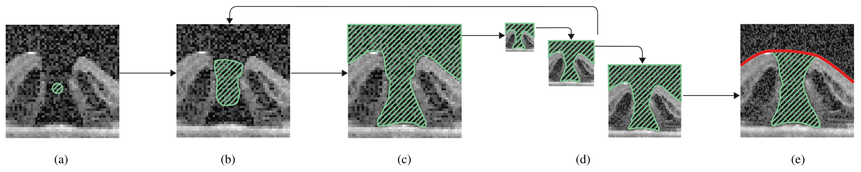
Given a 3D OCT image of a Macular Hole, this paper presents a fast and accurate fully-automated 3D level set segmentation method.
Citation
Please cite the article with:
@article{nasrulloh2017multiscale,
  author   = {A. V. Nasrulloh and C. G. Willcocks and P. T. G. Jackson and C. Geenen and M. S. Habib and D. H. W. Steel and B. Obara},
  journal  = {IEEE Transactions on Medical Imaging},
  title    = {Multi-scale Segmentation and Surface Fitting for Measuring 3D Macular Holes},
  year     = {2018},
  month    = {2},
  volume   = {37},
  pages    = {580-589},
  doi      = {10.1109/TMI.2017.2767908},
  ISSN     = {0278-0062}
}